Which Technology Would Be Best in Diagnosing a Broken Bone
Opens your nostrils with a nasal speculum. The bone density scan is usually recommended by the doctor when there is an undeniable loss in your weight fractured bone drop in hormone levels or intake of.
36 070 Broken Bone Stock Photos Pictures Royalty Free Images Istock
The x-rays length will depend on which part of the body the doctor is examining.
. This test checks the bone mineral density at a few places on your body and gives you a T-score The T-score compares your bones to those of a healthy 30-year-old. Digital x-ray Can be used to diagnose to a broken. Theres a New Method For Fixing Broken Bones Using Pieces of Other Peoples Bones.
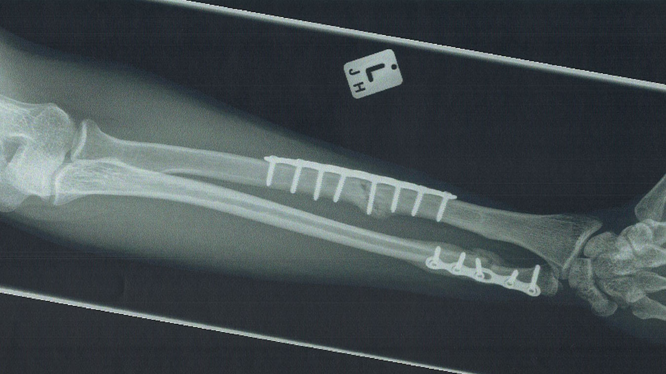
However it generally only takes a few minutes. But sticking a piece of metal inside the body can lead to various complications. A typical closed reduction is performed either by providing local anesthetic to the broken bone or a general anesthesia followed by a specific maneuver to attempt to realign the broken bone.
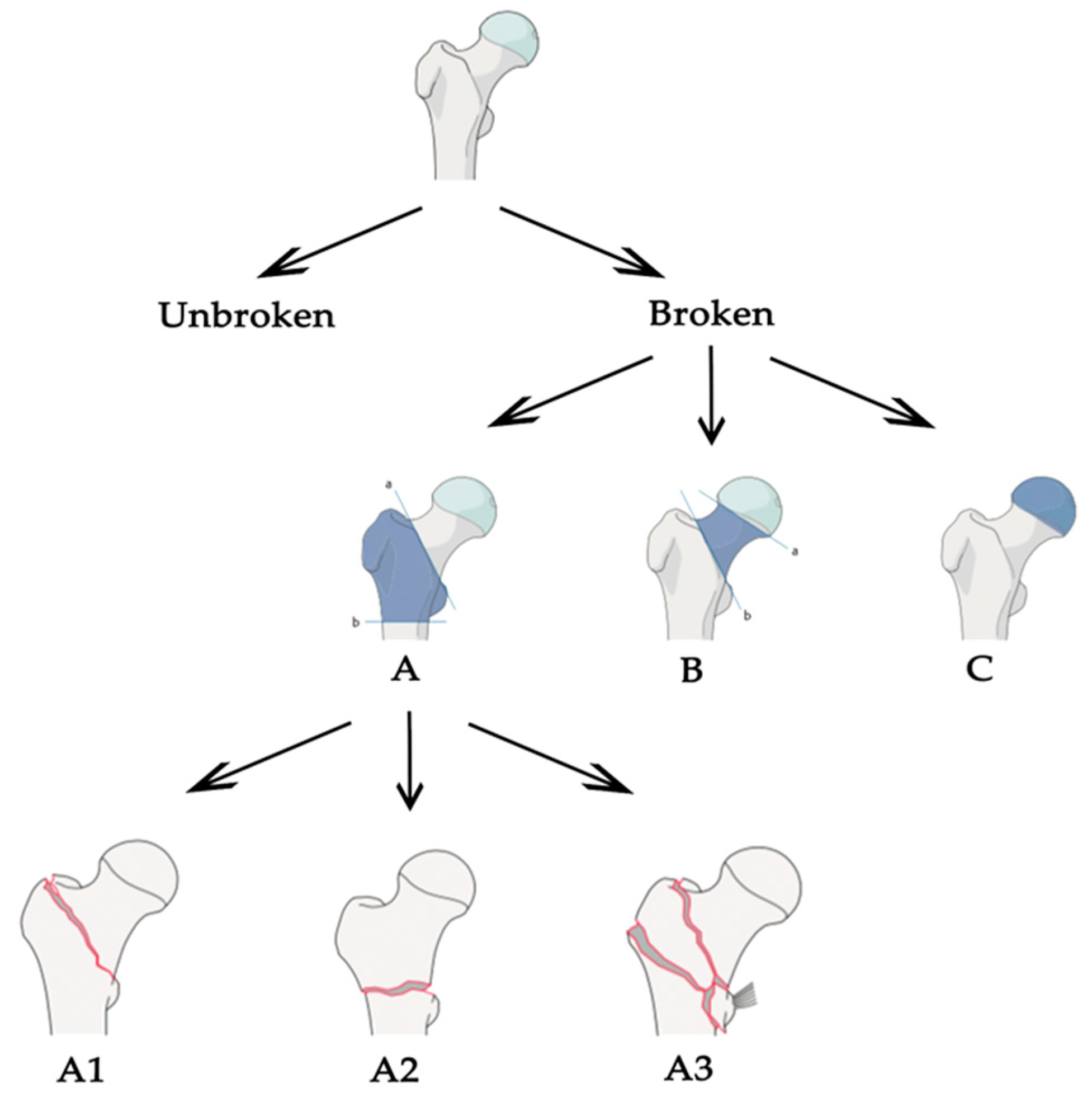
DXA is the best way to test bone mineral density. To diagnose a shoulder fracture an orthopaedic specialist orders advanced imaging tests to determine the exact location of your fracture and the severity of your condition. Bone fractures in various patterns and the types of broken bones will differ depending on the cause and nature of the fracture.
Radiography uses x-ray radiation to create images of the tissues organs bones and vessels that make up the human body. A CT scan shows details of the bones muscles fat and organs. These imaging tools let your doctor see inside your body to get a picture of your bones organs muscles tendons nerves.
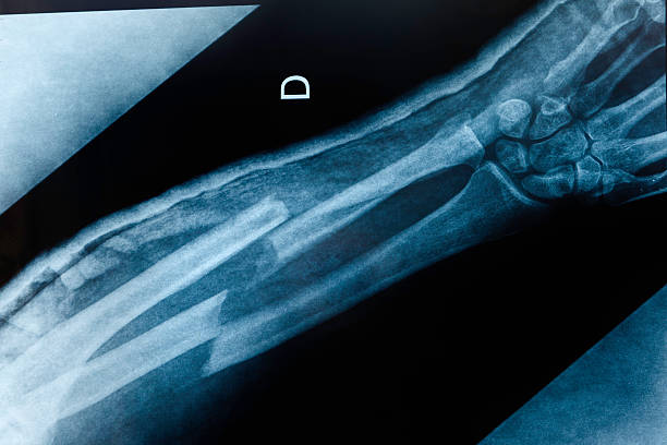
Most people are familiar with chest x-rays and also know that x-rays are the best way to diagnose broken bones. What to expect during an x-ray In this exam an x-ray machine sends individual x-ray particles through the body which are recorded as digital x-ray images on a computer. We offer the latest advancements in imaging tests and diagnostic tools to evaluate for shoulder fractures including.
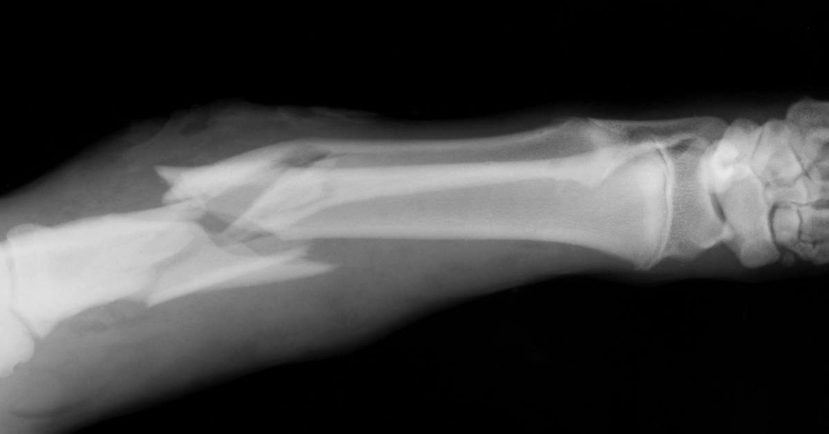
Your doctor will also splint your nose using packing in your nose and a dressing on the outside. A car accident will damage a bone differently than a disease which leaches out minerals and causes bone to rub together and rupture. Imaging tests such as X-rays are needed to accurately identify the nature of the broken bone and.
Using radio waves and a powerful magnet to produce detailed images of bone and soft tissues MRIs are much more sensitive than X-rays and can identify very small fractures and ligament injuries. Sometimes the only way to patch up a fracture is to use surgical screws that keep everything in place while the bone is healing. X-rays are the oldest and most commonly used medical imaging test.
Sometimes an internal splint is also necessary for a short time. Company called RegenTec has created a white powder that is designed to be injected into a person in order to speed up the healing process of broken bones. Diagnostic imaging techniques help narrow the causes of an injury or illness and ensure that the diagnosis is accurate.
After a closed reduction a splint or cast would be applied to hold the bones in the improved alignment while they heal. This is an imaging test that uses X-rays and a computer to make detailed images of the body. CT scans can often uncover rib fractures that X-rays might miss.
At Banner Health we are dedicated to supporting you in maintaining strong and healthy bones which is why we offer both conventional and digital x-ray imaging. A bone density test is different from a bone scan conducted to diagnose cancers fractures and other abnormalities in the bone. To evaluate damage to cartilage bone or other structures inside and around a joint MRI is the better choice.
The radiographer or x-ray technologist is the person who actually produces x-ray. But X-rays often have problems revealing fresh rib fractures especially if the bone is merely cracked. Now surgeons have come up with a novel type of screw.
However they have used them too for detecting types of cancer pneumonia and other developing conditions. CT scans are more detailed than general X-rays. X-rays CT Scans and MRIs.
Uses special instruments to help realign your broken bones and cartilage. Radiography X-Ray Technology Associate of Applied Science. X-rays are a good tool to visualize bone.
According to the Mayo Clinic there are four ways to diagnose a fractured rib. Doctors commonly use x-ray technology to diagnose broken bones but can also detect pneumonia types of cancers and other developing conditions. Radiologic technologists use X-rays sound waves and other tools of diagnostic imagery to create images of internal organs bones and tissues.
Osteoporosis can be found in more women than men. An X-ray can show injuries such as fractures infections arthritis and other changes. Doctors often use x-ray scans for diagnosing broken bones.
This technology takes X-rays from a variety of angles and combines them to depict cross-sectional slices of your bodys internal structures. The technology that would be best to diagnose a broken bone is by using an x-ray machine. These techniques include x-rays computed tomography CT scans and magnetic resonance imaging MRI.
You wont be able to just walk out of a hospital with a broken leg said Robin Quirk a professor at the University of Nottingham and co-developer of the technology. If youve ever broken a bone or had an ultrasound you were likely helped by a radiologic technologist. When it comes to testing for osteoporosis and your risk of broken bones the gold standard is dual X-ray absorptiometry or DXA.
Applied Sciences Free Full Text X Ray Bone Fracture Classification Using Deep Learning A Baseline For Designing A Reliable Approach Html
Sound Waves Are Almost As Good As X Rays For Detecting Broken Bones Techradar
Signs That You Have Broken A Bone San Antonio Emergency Room


Comments
Post a Comment